Friday, 1 June 2012

Pneumonia in Goats and Sheep

Introduction: Pneumonia, sometimes called goat shipping fever, is one of the most common problems encountered in sheep and goats today. Lambs and kids are particularly susceptible to pneumonia. It is a significant cause of decreased productivity and increased treatment costs. Early diagnosis and proper treatment are critical in controlling this problem.
Causative Agents: Most cases of pneumonia are caused by bacterial, viral, and parasitic infections. Pasteurella haemolytica and Pasteurella multocida are bacterial organisms carried in the respiratory tract of many normal animals. Most newborns are exposed to these organisms, but do not develop the disease because of natural resistance, a healthy environment, and ingestion of antibodies in colostrum that help control the infection. Viral agents such as parainfluenza-3 (PI3) are common in sheep and goats and can increase susceptibility to infection by causing inflammation of the respiratory tract. Certain infections of ovine progressive pneumonia (OPP) and caprine arthritis encephalitis (CAE) can cause pneumonia in sheep and goats. Other organisms, including Mycoplasma, Dictyocaulus (lung worms), and Eimeria can also cause lung problems.
In many cases, high humidity, dust, damp bedding, excessive heat, tight buildings with inadequate ventilation, and irritating gases such as ammonia compromise disease resistance and natural defense mechanisms in the sheep or goat, allowing pneumonia to develop. Weakness from a difficult birth, inadequate intake of colostrum, and other stresses contribute to the development of pneumonia in nursing animals. Often, a mild viral infection will occur, compromising the animal and allowing secondary bacterial infections to take place.
Clinical Signs: Young nursing animals that develop pneumonia commonly lose weight, become gaunt and lethargic, fail to nurse, and usually have a moderate fever. If the pneumonia remains undetected, serious lung damage will result and treatment will not be effective. Additional clinical signs include the following:
  1. Clear to yellow, runny to thick nasal discharge.
  2. Coughing and/or rapid breathing.
  3. Harsh lung sounds heard when listening with a stethoscope.
  4. Fever (temp. >103.5°).
Young animals that recover are susceptible to relapse during the feeding period and are more likely to suffer from heat stress and chronic cough. Coughing can lead to serious problems with rectal prolapse in feeder lambs.

Disease Transmission: Most of these infectious organisms are spread by direct contact with body fluids (saliva, nasal discharge, etc.) and fecal material. These problems can also be transmitted from one animal to another by contaminated hands, buckets, feeders, troughs, and equipment.
Diagnosis: Because lethargy and fever in sheep and goats may have several causes, a careful physical examination is required. In many cases, an exact (definitive) diagnosis is made by post-mortem (necropsy) examination. This list contains some key areas to help determine a diagnosis:
  1. The initial diagnosis can be made from general appearance and physical exam. The information on page C888 show regions of the lungs that should be listened to when using a stethoscope.
  2. Culture and sensitivity performed on nasal secretions or on samples taken at necropsy can help identify the specific cause of the infection.
  3. To accurately identify an infectious cause of pneumonia, a transtracheal wash performed by a veterinarian, with culture and sensitivity, may be necessary.
Treatment: Treatment must be based on early identification of affected individuals and depends on whether the cause is bacterial, viral, or parasitic. Fluid therapy, if practical, often helps the recovery rate. Producers should be sure that sick newborns are nursing or that they are provided supplemental milk via stomach tube. In serious outbreaks, it is often advisable to treat all exposed animals with a therapeutic dose of antibiotics for several days.
  1. Bacterial Causes -
    • Treatment with antibiotics such as penicillin, tetracycline, Albon, Gallimycin and even LA-200 may be considered. Like most bacterial infections, culture and sensitivity testing is recommended. See the antibiotic information in Section H.
  2. Viral Causes -
    • Treatment for all viruses involves treating the symptoms, not killing the virus. With this in mind, fluids, anti-inflammatory agents (Banamine), and antibiotics for secondary bacterial infections are recommended.
  3. Parasitic Causes -
    • Most parasites can be treated using ivermectin or doramectin. Routine de-worming will also help prevent the parasitic causes of pneumonia. Additional suggestions can be found on page B620.
Prevention and Control: Good management is the key to preventing respiratory problems. Producers with young animals that have pneumonia often have ventilation problems in the lambing/kidding and nursing facilities. Most commonly, the areas lack ventilation, resulting in high humidity and noxious gasses such as ammonia. In these conditions, the concentration of infectious organisms also rises. Well-ventilated (but cool) housing is probably ideal. Newborn animals should be dried off and then provided adequate colostrum intake. Weaker animals that fail to nurse should be provided colostrum via a stomach tube. Keep the bedding dry or newborns will chill and develop pneumonia. Do not skimp on bedding during the early nursing period.
Newborn animals should be provided supplemental heat only until they are dry and have nursed. Avoid overcrowding and do not keep the birthing areas too warm. Other than exposure to heavy drafts, healthy animals can tolerate cold fairly well. If electric fans are used, there should be at least four air changes per hour.
Parainfluenza-3 virus is a mild but common infection of young animals. Vaccination of young lambs (in problem flocks) with nasal IBR-PI3 vaccine at two or three days of age has helped reduce problems in some cases. The IBR vaccines should not be used in goats; there is a chance that the vaccine may actually cause this disease!
* If an animal has an infectious cause of pneumonia, it should be isolated from the rest of the flock/herd. No matter the cause of the pneumonia, caution should be used to prevent additional spread of the disease by contaminated clothing, hands, buckets, troughs, etc.

Pinkeye in Goats and Sheep

Pink eye is the lay term used to describe any number of diseases affecting the eye(s) of animals. The more proper name is infectious keratoconjunctivitis. Webster's Dictionary defines keratoconjunctivitis as "a combined inflammation of the cornea and conjunctiva."
Eye problemPink eye is an infectious and contagious bacterial disease of sheep, goats, and other animals. Though most common in the summer and in young animals, it may occur at any time of the year and in sheep and goats of any age. It occurs in all sheep and goat-raising areas of the world, though the primary causative organisms may vary.
Pink eye is caused by one of a number of different microorganisms. In the United States, the microorgansims most commonly associated with pinkeye in sheep and goats are Chlamydia psittaci ovis and Mycoplasma conjunctivae. The Chlamydia organism is the same organism that can cause enzootic abortions in ewes and does. Several other bacteria may play a secondary role in infections.
The causative organism(s) can be determined by a veterinary diagnostic lab from a scraping or swab of the conjunctiva. Depending upon the causative organism, pink eye can be contagious to people.

Symptoms

Pink eye tends to occur as an outbreak in a flock or herd. The causative organism is commonly introduced via purchased sheep or goats. The microorganisms that cause pink eye are widely distributed and may persist in resistant, carrier animals. Spread occurs via direct contact. Sheep and goats raised under intensive conditions are most commonly affected.
Lamb with eye problemWhen sheep and goats eat out of the same trough, they can easily transmit or contract the disease. Overcrowding and poor ventilation contribute to the spread of the disease. Dry, dusty conditions and irritants such as flies tend to predispose or exacerbate the disease. Flies or dust can carry the bacteria to the eye.
Whatever organism is responsible, the symptoms are similar. Affected animals blink repeatedly and have an aversion to bright sunlight. The side of the face below the eye may be wet due to tearing. Upon close examination, the membranes of the eye appear red and inflamed. The eyes become cloudy or opaque. An ulcer may develop. The condition is painful and may affect one or both eyes. Pink eye can cause temporary blindness in affected animals; permanent blindness in severe cases.
Sheep and goats rarely die from pink eye. The cost of the disease is associated with treatment costs (medicine and labor) and production losses, as affected animals may have difficulty finding food and water. Animals that suffer temporary blindness may require hand feeding or watering to prevent production losses.

Treatment

Affected animals should be isolated from the rest of the flock to prevent spread of the disease. They should be housed in a clean, dry, comfortable, and shady place.
Pink eye is usually treated with any number of antibiotics that are injected into the body or placed directly in the eye. The most common treatment is to apply terramycin ointment to the affected eye(s) two to four times per day. Some veterinarians advocate the use of intramammary mastitis tubes for the treatment of pink eye. As with the terramycin ointment, the antibiotic is applied directly to the eye.
Crusty eyeOintments are usually more effective than powders or sprays. They are less irritating to the already inflamed eye. Eye drops are easier to administer than ointments. Before applying medicine to the eye, the animal's face should be cleaned and the debris around the eye(s) should be removed. Surgical gloves should be used when affected animals are handled.
Sometimes, subconjunctival injections of penicillin are given or the antibiotic is dropped into the eye. When it is not practical to treat individual animals repeatedly, antibiotics may be injected systemically. Long-acting oxytetracyclines (e.g. LA-200®) are most commonly used. The additition of oxytetracycline to be the feed may be beneficial. Tylosin (Tylan®) is also effective against the causative organisms of pink eye.
Because most of these antibiotic treatments are not FDA-approved to treat pink eye in sheep and/or goats, veterinary advice should be sought. Extra-label drug use requires veterinary approval, even if the drugs can be purchased over-the-counter or via mail order. Subconjunctival injections should not be attempted by lay people.
Despite intensive efforts, treatment may have little effect on the course or severity of the disease. Pink eye is similar to sore mouth (orf) in that the disease is usually self-limiting and the majority of affected animals will clear up without treatment, usually in a week to 10 days. Severely affected animals may take longer to recover. Recovered animals have resistance for varying lengths of time. It is possible for them to become reinfected, as acquired immunity is not strong or long-lasting.

Prevention

Sheep with eye problemThere is no vaccine to prevent pink eye in sheep and goats. Because the microorganisms that cause pink eye in cattle (Moraxella bovis) are different from the ones that cause pink eye in sheep and goats, the vaccine used to prevent pink eye in cattle will not prevent pink eye in sheep or goats.
The best way to prevent pink eye is to maintain a closed flock or herd. Do not purchase animals from public auctions. Isolate new livestock for at least 30 days. Show animals should also be isolated upon returning to the farm, as pink eye is common at fairs and expositions.
Dust and fly control will aid in the control and spread of pink eye. Protection from sunlight should be provided. Complete disease eradication is difficult because the organisms that cause pinkeye are widespread and may persist in carrier animals.

Milk Fever in Goats, Sheep and Cows

 Goat's/Sheep With Milk Fever
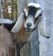
If your doe is shivering like she’s cold, don’t assume she needs a sweater. Your first guess should be milk fever.
Hypocalcemia, commonly known as milk fever, is a calcium deficiency found in goats and cattle. Most often seen in newly freshened does, milk fever can also be found in goats who are preparing to kid, are from high production lines and even in those not being milked at all.
The most common signs of milk fever are shivering during or after milking, wobbly legs – the goats seems off balance and may run into things, and general lethargy. Other symptoms can include poor appetite, poor milk production, decreased body temperature, rumen dysfunction, weakness or an inability to stand. Left untreated, milk fever can cause death.
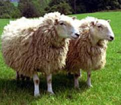
Goats with milk fever should be immediately treated with supplemental calcium. Calcium Gluconate given orally, Tums, high calcium food stuffs such as molasses, parsley, bok choy, tofu, kale and alfalfa can be added to the goats diet. There are also calcium supplements on the market specifically for treatment of calcium deficiency. If the doe has a severe case (doe is down), a commercial or veterinary product is the way to go. If the doe is shivering but otherwise appears healthy, a more natural approach can be taken but it is imperative that the calcium level be returned to normal as soon as possible.


Cows with Milk Fever

Milk fever is a disorder mainly of dairy cows close to calving. It is a metabolic disease caused by a low blood calcium level (hypocalcaemia). Between 3% and 10% of cows in dairying districts are affected each year, with much higher percentages occurring on some properties. Jersey cows that are mature and fat and graze lush, clover dominant pasture before calving are most susceptible.
Losses are due to deaths (about one in 20 affected cows dies), a reduction in the productive lifespan of each affected cow of about three years, and reduction in milk production following each milk fever episode, as well as costs of prevention and treatment.

Signs
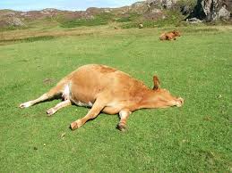
In typical cases cows show some initial excitement or agitation and a tremor in muscles of the head and limbs. Then they stagger and go down to a "sitting" position, often with a 'kink' in her neck, and finally lie flat on their side before circulatory collapse, coma and death.
A dry muzzle, staring eyes, cold legs and ears, constipation and drowsiness are seen after going down. The heart beat becomes weaker and faster. The body temperature falls below normal, especially in cold, wet, windy weather.
These signs are due mainly to lowered blood calcium levels. Sometimes there are additional signs due to complicating factors. Bloat is common in cows unable to "sit up" because the gas in the rumen is unable to escape. Pneumonia and exposure may affect cows left out in bad weather.

Causes

About 80% of cases occur within one day of calving because milk and colostrum production drain calcium (and other substances) from the blood, and some cows are unable to replace the calcium quickly enough. High producers are more susceptible because the fall in their blood calcium level is greater. Selecting cows for high production may, therefore increase the problem with milk fever. Some individual cow families or breeds (for example, Jerseys) are more susceptible than others.
Age is important. Heifers are rarely affected. Old cows increase in susceptibility up to the fifth or six calving because they produce more milk and are less able to replace blood calcium quickly.
The feeding management of dry cows in the 2 weeks before calving is very important, because it affects both the amount of calcium available to replace blood calcium and the efficiency with which the available calcium can be used.
When the amount of calcium in the diet is greater than is needed, the efficiency of absorbing calcium from the intestine and the efficiency of transferring calcium from the skeleton both become very sluggish and the chance of milk fever is greatly increased.
Also, grazing pastures in Southern Australia winter and spring results in alkaline blood which creates conditions unfavourable for the availability of calcium in the body and predisposes the cow to milk fever. Feeding hay prior to calving and restricting access to green feed results in acidic blood which favours calcium mobilisation from bone and improves calcium absorption from the intestines, both of which are important factors in preventing the occurrence of milk fever.
Fat cows are at a greater risk than thin cows. This is partly because their feed and calcium intake has been higher and partly because fat cows produce more milk at calving time.
Some cows get milk fever several days or even weeks before or after calving. This is usually due to the feed, especially the dietary calcium, being insufficient to meet the heavy demand due to the rapidly growing foetus or milk production in early lactation.
In early lactation, cows should receive as much calcium as possible, and clover-dominant pasture are therefore desirable. They will help to prevent grass tetany as well as milk fever.

Treatment

Treatment should be given as soon as possible. Use 300 ml, or more, of a 40% solution of calcium borogluconate or, preferably, a combined mineral solution such as "three-in-one" or "four-in-one". Often 600ml may be required.
The combined solutions contain additional ingredients such as magnesium, phosphorus and dextrose (for energy), which may also be at low levels in the blood while cows have milk fever.
Packets of solution together with an injection kit are best kept on hand for emergencies. All equipment should be kept sterile to avoid abscess formation at the site of injection.
Injection of the solution by farmers should be in several places under the skin on the neck or behind the shoulder, unless the cow is in a coma or there are other reasons for desiring a quick response.
Injection into a vein should be left to a veterinarian as it can cause sudden death if not carried out properly. Veterinary assistance is also advisable if there is not a quick response to treatment, because other problems may also be present.
Cows that are "flat out" should be propped up into a normal resting position to relieve bloat. If weather conditions are bad, or the response to treatment is slow, transfer the cows to shelter to prevent exposure and other complications. Provide feed and water. Rugging helps.
Some cows that have been comatosed may have regurgitated and inhaled rumen content into the lungs. If there is ruminal material around the nose one should be suspicious that this may have happened and intensive antibiotic treatment should be commenced as soon as possible as inhalation pneumonia is often fatal.
Recovered cows should not be milked for 24 hours; then the amount of milk taken should be gradually increased over the next 2-3 days.

Prevention

Management of the diet can be a valuable aid preventing milk fever. Cows should be kept on a low calcium diet while they are lactating (dry). This stimulates their calcium regulatory system to keep the blood levels normal by mobilising the body stores of calcium from the bone. When the demand for calcium increases as calving, calcium can be mobilised much more rapidly from bone than the feed, therefore preventing milk fever.
With cows at greater risk - Jersey cows of mature age and in forward to fat condition - green feed should be restricted and plenty of hay fed for at least 1-2 weeks before calving. Neither should contain a high percentage of clover or capeweed.
If it is necessary to improve the body condition of cows in order to improve milking performance, feeds high in energy but low in calcium may be used, for example cereal grain or oaten hay. Cereal grain is also high in phosphorus content, and this is of additional value.
Cows close to calving should be kept in a handy paddock to enable frequent observation and early detection of milk fever. On the point of calving, and afterwards, the available feed and calcium should be unrestricted. Calcium feed supplements may be helpful at this point, but should not be given earlier.
Where dietary management is inadequate, other methods are sometimes used. Vitamin D3 given by injection 2-8 days before calving may be useful. As the calving date is often difficult to predict, repeated treatments are sometimes necessary.
A common treatment used to prevent milk fever is the injection of calcium borogluconate just before or just after calving. Some cows are given more than one treatment. This is quite successful because the calcium provides a reservoir to increase blood calcium just at the time it is needed for milk and colostrum. The danger is that it may not last long enough and milk fever may still occur before the calcium-regulating mechanism of the cow is working efficiently.
Drenching cows with Unimix on the day before and then twice daily for 1 to 2 days after calving has considerably reduced the incidence of milk fever in some herds where other methods alone have been unsatisfactory. Unimix is a registered product containing a mixture of calcium and magnesium.
Cows that have required injections to treat milk fever will benefit from a drench of Unimix to help prevent relapses.

Mastitis in Goats

The organisms infecting the udder of goats are similar to those in cows. Coagulase-negative staphylococci are generally the most prevalent and can cause persistent infections that result in increased cell counts and low-grade mastitis with some recurring clinical episodes. The level of infection and incidence of mastitis due to Staphylococcus aureus tends to be low (<5%), but can result in persistent infections that do not generally respond to therapy. Streptococcal intramammary infections can occur in both subclinical and clinical cases, but are usually much less frequent than in cattle. Streptococcus agalactiae is not a common pathogen of mastitis in does.
Mycoplasma infections, primarily M mycoides (large colony type) and M putrefaciens , sometimes cause serious outbreaks of mastitis in goats (see also Contagious Agalactia and Other Mycoplasmal Mastitides of Small Ruminants: Introduction). The latter also causes septicemia, polyarthritis, pneumonia, and encephalitis, together with serious disease and mortality in suckling kids. M capricolum has also been reported to cause severe mastitis in goats and infection in kids. Does usually recover in ~4 wk.
As with cows, gram-negative organisms cause intermittent infections that may be severe but are usually self-limiting. Arcanobacterium (Actinomyces) pyogenes sometimes produces multiple, nodular abscesses.
Does can also exhibit signs of mastitis from caprine arthritis and encephalitis ( Caprine Arthritis and Encephalitis: Introduction) and ovine progressive pneumonia ( Progressive Pneumonia) secondary to systemic infection. Agalactia is common, as is a hardening of the udder from fibrosis.
Programs for diagnosis, control, and treatment of bacterial mastitis in goats are similar to those in cows. However, monitoring subclinical mastitis with SCC in does is difficult due to poor discrimination between infected and noninfected animals, especially in the later stages of lactation. This is partially because a higher proportion of cells in goat milk are epithelial in origin as compared with cow milk. As lactation progresses, shedding of epithelial cells into milk increases, thus SCC >1,000,000 cells/mL are common in uninfected does in late lactation. Proper milking procedures and good environmental sanitation are needed to reduce the prevalence and spread of infection. Chronically infected goats should be culled, as should goats with M mycoides infections and those that do not recover from M putrefaciens or M capricolum infections.

Pregnancy Toxemia and Ketosis in Does (Sheep and Goats)


Pregnancy toxemia and ketosis are the result of the high carbohydrate (energy) demand of multiple fetuses in late pregnancy. The kids require an increasing amount of carbohydrates the last trimester. Does bearing twins have a 180% higher energy requirement than those with just a single fetus. Does carrying triplets have a 240% greater energy requirement. When this demand exceeds the supply, fat is metabolized into glucose. The metabolic needs of the kids are met at the expense of the dam; this is what causes the ketotic condition. To complicate matters, multiple fetuses produce more waste products, which leads to the doe becoming toxic if she does not flush them from her system.
Risk Factors for Pregnancy Toxemia
  • Multiple fetuses
  • Poor quality of ingested energy
  • Dietary energy level
  • Environment
  • Genetic factors
  • Obesity
  • Lack of good body condition or high parasite load
  • Confinement - lack of exercise

Toxemia and ketosis are typically seen in does that are overweight and get little exercise. Under weight animals that are fed a poor quality feed are also candidates for toxemia. Look for does at the bottom and top of the pecking order. These does may be getting to much or not enough feed. Does should be in good body condition, and not overly fat when bred. They can be maintained on good roughage or forage during the first 100 days of pregnancy. During the last trimester the doe should gain approximately 1/2 lb. per day. The doe must intake enough carbohydrates to supply the demand of the growing fetuses and to keep her alive and functioning also.
I also believe that we are seeing an increase in toxemia this year due to the extended drought conditions. With these conditions the quality of the feed changes, the browse is limited, and the animals do not receive the vitamins and minerals that they get naturally from high quality feeds and browse. Extremely wet conditions, especially if following a prolonged drought can also cause a dramatic and quick increase in the worm load, and cause the doe to drop enough body condition to become a candidate for toxemia.
When there is a decrease of glucose levels in the doe's brain, they tend to lay down, become sluggish, and show a loss of appetite. They may get stiff, and walk with a staggering gait. Swelling (edema) of the lower limbs is not uncommon. Some does may also grind their teeth. Keto acidosis is also common during toxemia and needs to be treated also. As the disease progresses, the neurological systems become compromised due to lack of glucose. Blindness, stargazing, tremors, aimless walking, ataxia (uncoordinated staggering gait), are seen and eventually the doe becomes comatose. At this stage the fetuses succumb and release toxins that send the doe into endotoxic shock, and death. Does that survive toxemia need to be watched for dystocia, and lactational ketosis.
Diet should include high quality roughage and increased concentrates. At fist sign of decreased appetite, or unwillingness to rise, managers need to be wary. Exercise should be offered and forced if necessary. Some type of high-energy supplement needs to be given to keep the doe from coming ketotic. The carbohydrate (energy) level of the diet needs to be increased. This can be accomplished by adding corn, fresh alfalfa hay, or a soybean supplement to the diet. Increasing the protein does not necessarily increase the energy level.
High Energy Supplements
  • Propylene Glycol
  • Nutri-drench
  • Dextrose
  • Environment
  • Magic (1 part Molasses, 2 parts Kayro, 1 part Corn Oil)
  • Glucose IV
B Complex, thiamin, probiotics, and penicillin should also be given as support therapy. 3cc B Complex and 500mg thiamin should be given twice daily. Probiotics should be given at least daily, and a large initial dose of penicillin followed by substantial doses for 5 days total should also be administered. This also helps prevent primary and secondary pneumonia.
If the kids are within 7 days of due date, and the doe does not respond immediately to treatment, giving 20-25mg dexamethasone can induce labor. Labor will begin within 48 hours. Dexamethasone is preferred over hormonal induction of parturition because of its beneficial stimulus to appetite. Also, dexamethasone may also prepare the lungs of marginally immature kids increasing their chances of survival during labor or cesarean. Valuable does that fail to respond within 24 hours should have a cesarean immediately. Even with surgery and fluids, prognosis is poor for the survival of both the doe and kids.
The key to treating toxemia is catching the subtle symptoms promptly. Fist concern is to hydrate the doe and get enough sugars to the brain to get if functioning properly. We have been successful treating does with 50% Dextrose solution and magic. We give 20cc magic two to four times daily at first signs of toxemia. If the doe is completely off feed or becoming lethargic we give 4 ounces 50% dextrose (1/2 dextrose, ½ water) with 3cc B Complex, 500mg thiamin, 50cc Calcium Glutamate (orally) and probiotics. The dextrose solution is repeated every hour until the doe is standing, drinking, and urinating. We have also found it helpful to give water (drench if necessary) at every treatment. Once these does quit moving, they don't get up and drink, as they should. We give one pint of water with every treatment.
Be cautious given electrolytes or products that are high in sodium. Although in a pinch they will work to hydrate the animal, you don't want to give long term anything that has a lot of sodium in it. That is why we use the dextrose instead of oral electrolytes. You need to make sure that the doe urinates, giving a mild diuretic such as cranberry juice may also be helpful. The does need to flush the toxins from their system.
When the doe starts to respond we start decreasing the treatment intervals. Treatment is dropped to 4 times a day, and if the doe is back on her feed and eating well, we just give the magic mixture two to three times daily. The doe may scour the first couple of days & Pepto-Bismol or kaopectate can be given to relieve the dietary scours. Don't be terribly concerned if the doe scours it is caused by the drastic dietary changes and will clear up as the doe continues to eat.
Propylene glycol can be given orally at the rate of 4 ounces, 4 times daily. Nutri-drench can be substituted for propylene glycol, and is easier to dose orally - it doesn't taste quite as offensive to the doe. Intravenous glucose (25-50 grams - in a 5 or 10% solution), and B Vitamins, can also be given. If probiotics are not available give baking soda orally to prevent acidosis these need to be given at least once a day. Dextrose (50%) can be given IV in a single 60-100ml dose, followed by a 5% dextrose solution in an electrolyte drip. Be careful giving commercially prepared and oral electrolytes, they are usually high in sodium.
Ketoacidocis is precipitated by metabolism of fat. When the doe is not consuming an adequate amount of carbohydrate, that she metabolizes fat to make glucose. The byproduct of this is ketones, which must be secreted by the kidneys. The doe must have a fluid intake to allow this to happen. That is why plain water is given, drenched, or tubed, along with the 50% glucose, and other support therapy. When enough glucose is present, fat is not metabolized, and the body has time to get rid of it. As this happens, the doe will become more alert, and start to eat and drink on her own. This is a vicious cycle, and difficult to break. If caught early, and treated concienciously, the cycle can be broken. The doe must be monitored carefully until the kids are born, condition can change quickly and needs to be responded in a timely fashion.

How to trim your Goat's/Sheep's hooves (Prevent Hoof Rot)

Tools The first thing to consider is your selection of tools.
    From left to right are
  • Rasp - used to flatten the bottom of the hoof after trimming. The raised end, at the bottom in this photo, rests in the palm of your hand. There is a slight indentation in the top of the other end where your forefinger and middle finger sit. The tool is operated by pressing it down and forward, toward the end with your fingers.
  • Trimmers - select a sharp pair. We use the "orange" handle ones from Caprine Supply.
  • Knife - a *very* sharp knife. Works well in reaching those areas your trimmers can't get to. Also good for flattening the sole and for removing embedded rocks and thorns.
  • Brush - it helps if you brush off the junk so you can see what you're doing.
  • Blood Stop Powder - If you're careful and lucky you won't need this.

First, remove any dirt from the outside of the hoof and the dig dirt, mud, and nanny berries from the bottom of the hoof. A good tool for this is a hoof pick (not shown) or a nail. You can use the tips of your trimmer but by doing so you'll cut their life in about 1/3.
Hoof trimming takes time and is physically stressful for both you and the goat. It's very important that a comfortable position is established. We prefer having the goat secured on a fitting stand or other raised platform (though in my younger more flexible days I sometimes left the goat on the ground and just clipped it to a fence). Fitting Stand

First trin the toe The first cut on a badly overgrown foot like this one is to nip away the front of the nail. This makes it easier to cut the sides of the nails by opening a slot for you to fit the trimmers into.


Once the front tip of the nail is removed it's easy to insert the trimmers to cut the sides of the nail. You only need to cut it back even with the sole (also called the "frog") at this point. Trim the nail

Continue to trim the sides around one toe and then across the heel. Repeat the process on the other toe.

The frog and heel can be trimmed flat with either a knife or the rasp. When using the knife cut from the toe toward the heel. When using the rasp go from the heel to the toe.
This is the point where you might need the blood stop powder. Depends on how deep you go with the knife. Trim off a very thin slice with each stroke - a good rule is to stop when you see pink. Same thing goes for the rasp.
We stopped before we got this hoof completely flat. Note the dark brown areas on the sole.

The nail, sole, and heel have been trimmed parallel with the growth bands around the outside of the hoof and we were seeing pink on the sole. We'll come back in a couple of weeks and use the rasp and/or knife to finish the flatening process.

Hoof Rot in Goats

The first sign of hoof rot is usually lameness; the goat is limping or holding the hoof off of the ground while standing. Many time more than one hoof on an animal will be infected. Inspection of the hoof will show separation of the horny portions of the hoof from the soft tissue. When the horny surface is trimmed away you will notice a foul-smelling paste in the area.
Causes

      
The bacteria that causes hoof rot (Bacteroides nodosus and Fusobacterium necophorum) work together to cause hoof rot and can be found in the ground just about anywhere goats are raised. These bacteria are anaerobic which means that they can live in free air for only a short time. Wet, muddy ground is a particularly good place for the bacteria to hide.
Insufficient zinc in the diet can result in deformed hooves which crack and provide bacteria a way to enter the hoof.
Overgrown hooves will allow the horny surface to curl inward over the sole of the hoof. This provides an excellent area for bacteria to grow. Routine hoof trimming is vital. My article on hoof trimming will be displayed at the end of this article.
Some breeding lines have a higher rate of hoof problems than others. Include the ancestors' hoof health in your evaluation criteria for prospective purchases.
Treatment

      
Individuals with hoof rot
Remove any foreign objects such as nails, small pebbles, etc., from the hoof. Pay particular attention to the space between the horny surface and the soft tissue.
Perform a complete hoof trimming then trim away any horny "toe nail" from the affected area. Thoroughly clean the area to remove all traces of the "pus". Consult your vet for their recommendation of medication. Some vets will advise penicillin or oxy-tet injections while others feel that topical (direct) medication such as Kopertox®, merthiolate, or mercurichrom applied to the infected area is better or perhaps should be administered at the same time as the injectable medication.
Quarantine the infected goat. It's ok to keep a group of goats together that have the same disease - just don't let them out with the rest of the herd and don't let "clean" animals in with them.
The rest of the herd
A preventive measure that seems to work is the use of a daily footbath of zinc sulfate. Mix 8 pounds of fine zinc sulfate with 10 gallons of water. Stir in 1 cup of laundry or dish soap to reduce the surface tension of foreign matter on the hooves and allow it to fall off more easily. For large number of animals you will need to increase the amount of each ingredient proportionately.
Set it up so the herd has to pass through the footbath to get to feed or water. They will be reluctant initially to walk through it but quickly adapt. The solution must be replenished and changed periodically. Good quality drinking water and salt must be made available so the goats are not forced to drink the foot bath solution.
Replenish or replace the solution as needed.
Hoof rot is expensive in the form of increased labor and in money. Animals that cannot walk correctly because of hoof rot will not grow as well as their herd mates and have a decreased value to you as a breeder and to prospective meat market buyers. Such animals will also have a hard time escaping predators. Does will have difficulty standing to be bred and bucks may not be able to mount the doe if a rear hoof is badly infected.